Human Imaging Research Office
The University of Chicago Biological Sciences
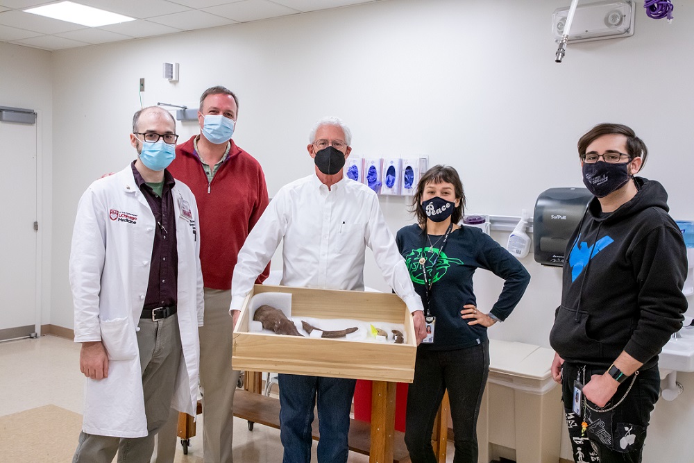
HIRO lends a hand and helps scan SUE the T. Rex's arm!
Researchers at the Field Museum of Natural History are hoping to finally determine how Tyrannosaurus Rex used its arms, and the Human Imaging Research Office was able to help! Staff from the HIRO worked with UChicago Medicine Radiology to perform CT scans on the right arm of SUE the T. Rex, one of the world's largest and most complete T. Rex specimens. The research team at the Field Museum, led by paleontologist Jingmai O'Connor, enlisted the help of radiologist Christopher Straus, M.D. and the HIRO to perform extremely detailed high-resolution CT scans of SUE's arm and shoulder. These CT scans will be used to create 3-D models of the individual bones so Dr. O'Connor's team can model the biomechanics of T. Rex's arm. The project was covered extensively in the local media by outlets like the Chicago Sun-Times, CBS2 & WBBM, ABC7, and FOX32.
For more information, check out the media coverage below:
- ABC7 News - CT scans of SUE the T. Rex hope to reveal more about dinosaur's famously short arms
- Chicago Sun-Times - Field Museum’s Sue lends a hand to study of T. rex’s short arms
- UChicago News - SUE the T. rex’s right arm undergoes CT scan at UChicago Medicine
Health Imaging - Experts hope CT can help unlock age-old question: Why were T. Rex arms so short?
The research team holds SUE's arm while in the CT scanner suite at UCM's Center for Care and Discovery.
From L to R: Nick Gruszauskas (UCM HIRO), Christopher Straus (UCM Radiology), Bill Simpson (Field Museum), Jingmai O'Connor (Field Museum), Ryan Davila (Field Museum).
Photo credit 2022 Jordan Porter-Woodruff/UChicago Medicine
News item posted on 2022-12-08 17:00:00 -0500.